Как делать ставки в БК Леон: регистрация, идентификация, процесс игры в интерфейсе букмекерской конторы
Словарь беттинга
Автор РУСбеттинг На чтение 5 мин Опубликовано 24.01.2022
Букмекерская компания «Леон» принимает интерактивные ставки на сайте leon.ru. Игрокам доступна как полная версия сайта, так и его мобильная версия. Обратите внимание: играть на официальном сайте букмекерской конторы могут только граждане РФ. Валютой игрового счета является только российский рубль.
Шаг 1. Регистрация на сайте букмекерской конторы «Леон»
С 1 октября все вводы и выводы средств проходят через Единый ЦУПИС, и одного ID там достаточно, чтобы играть во всех легальных букмекерских конторах России.
Чтобы начать делать ставки в букмекерской конторе «Леон», перейдите на сайт www.leon.ru и нажмите на кнопку регистрации в правом верхнем углу экрана.
После этого необходимо заполнить появившуюся форму: ввести номер мобильного телефона, адрес электронной почты, дату рождения и придумать пароль не менее чем из семи символов.
Регистрационная форма содержит ссылки на условия и правила, используемые букмекерской конторой Леон. Прежде чем принимать, рекомендуем изучить их: у разных букмекерских контор разные правила приема и расчета ставок.
Для завершения процедуры регистрации необходимо принять правила БК «Леон» и дать разрешение на обработку персональных данных. Кроме того, игрок должен подтвердить достоверность предоставленной им информации и принять условия Единой оферты ЦУПИС. Установите соответствующий флажок. После этого нужно просто пройти капчу и нажать кнопку «Регистрация».

№ 5 20500 ₽ Играть
Шаг 2. Идентификация личности в БК «Леон»
Согласно законодательству РФ, для игры на сайтах российских букмекерских контор необходимо иметь учетную запись в учетном центре интерактивных букмекерских переводов, которым также является ЦуПИС. Центр учета интерактивных букмекерских трансферов выступает посредником между игроком и букмекерской компанией, отвечая за денежные операции между сторонами.
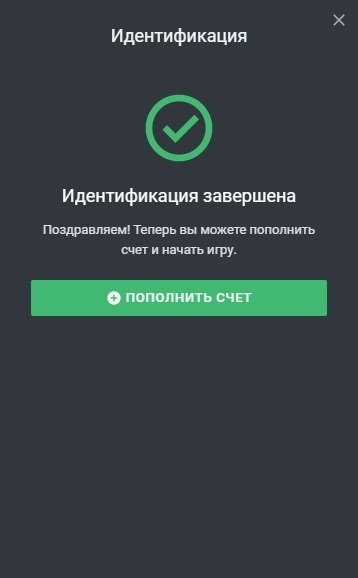
Сразу после регистрации на сайте leon.ru указанный игроком номер телефона будет проверен. При подключении верифицированного аккаунта в Едином ЦУПИС пользователь сможет сразу же начать отыгрывать ставки; идентификация будет завершена автоматически.
Если у клиента нет верифицированного аккаунта в Едином ЦУПИСе, букмекерская контора предложит несколько вариантов прохождения упрощенной идентификации онлайн:
- ручной ввод паспортных данных клиентом, не нужно подавать документы;
- идентификация через портал «Госуслуги»;
- идентификация путем загрузки фотографии паспорта (только главная страница).

Упрощенная онлайн-идентификация удобна, но имеет определенные ограничения, которые не подойдут любителям высоких ставок.
При упрощенной идентификации личности максимальный размер одной операции (пополнения счета/снятия средств) составляет 60 000 рублей, а максимальный месячный оборот средств — 200 000 рублей.
Снять все лимиты можно после прохождения полной идентификации. Вам придется сделать это лично, предъявив паспорт:
- в магазинах «Связной» (стоимость услуги 300 рублей, артикул 231695);
- в отделениях платежной системы CONTACT (стоимость услуги 150 рублей, пункт QONR).
Шаг 3. Пополнение счета
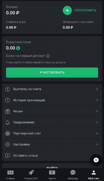
Для совершения первых ставок необходимо пополнить игровой счет на сайте букмекерской конторы «Леон». Для этого нажмите на плюсик рядом с балансом в правом верхнем углу экрана, либо перейдите на экран пополнения через профиль.

Для пополнения счета вы можете использовать любой из предложенных способов оплаты:
- банковские карты (VISA/MasterCard/Maestro/Cirrus/Мир»);
- электронные кошельки (QIWI, ЮМани, ЦУПИС, WebMoney);
- мобильные платежи;
- Apple Pay, Google Pay, Samsung Pay;
- «Альфа-Клик».
После выбора способа оплаты откроется страница, где игроку будет предложено ввести сумму для зачисления.
Средства зачисляются на счет в «Лионе» моментально. Минимальная сумма для пополнения игрового счета в этой букмекерской конторе составляет 100 рублей.
№ 5 20500 ₽ Играть
Шаг 4. Игра в букмекерской конторе
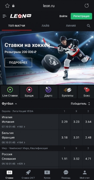
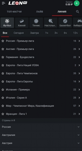
На главной странице покупателя встретит раздел «Топ-матчи», в котором собраны самые интересные предстоящие события. Вверху есть отдельные столбцы: «Основные матчи», «Лайв» и «Линия».
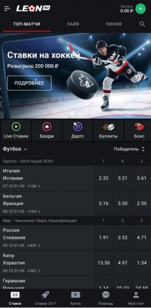
В строке названия турниров указаны в алфавитном порядке, но самые популярные соревнования размещены в начале.
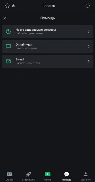
В разделе «Помощь» вы также можете найти информацию о том, как делать ставки.
Ставки принимаются в два клика: сначала игрок выбирает свой вариант ставки, затем подтверждает в окне купона, которое открывается внизу экрана.
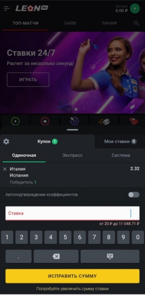
Расчет стандартных ставок по стандартным котировкам (результат, итог) происходит быстро, иногда даже во время мероприятия.
История транзакций находится в купоне. Игрок может просматривать операции по счету за последние 15 дней и за предыдущие два месяца.

Шаг 5. Вывод денежных средств
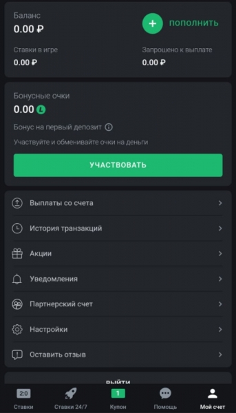
Вывод заработка осуществляется через меню «Платежи по счету» в профиле клиента. При нажатии игрок должен выбрать способ вывода и ввести желаемую сумму вывода.
Букмекерская контора предупреждает, что может потребовать вывод средств с помощью платежной системы, с помощью которой игрок пополнял баланс, однако четкого ограничения в правилах нет.
Для вывода средств можно использовать:
- банковские карты (VISA/MasterCard/Maestro/Cirrus/Мир»);
- электронные кошельки (QIWI, ЮМани, ЦУПИС);
- банковский счет.
При создании заявки на вывод букмекерская контора напоминает, что при выводе выигрыша более 15 000 рублей Леон выступит в роли налогового агента и удержит налоги с выигрыша (13% от чистого выигрыша). Чтобы этого избежать, компания просто ограничила размер единовременной выплаты, ведь при получении выигрыша в размере до 15 000 игрок несет ответственность за уплату налога.
Подробнее о налогообложении выигрышей можно прочитать в статье Налогообложение интерактивных ставок.
После выполнения заявки на вывод средств контора букмекерской конторы «Леон» отправляет СМС на телефон игрока с кодом, подтверждающим запрос.
По информации службы поддержки компании, в некоторых случаях при выводе средств БК «Леон» может потребоваться дополнительная проверка личности — букмекерская контора уведомляет об этом по электронной почте.
- Для тех, кто хочет играть на мобильном устройстве, у БК «Леон» есть как приложение для Android, так и приложение для iOS.
мутная контора, если в например в лайв событии счет 2:0 или вешают замки или на выигрывающего прочерки и сможешь поставить только заведомо проигрышную ставку, кстати параллельно смотрел в другой бк это событие — там вплоть до 1.00001 можно поставить
Главное правило всех букмекеров – любой бонус должен быть отыгран. В БК Леон это делается следующим образом:
БК Леон раздает клиентам фрибеты за депозит до 17 февраля 2023 года.
Flashscore RU FlashScore Russia · Спорт
Если пари клиента проиграет, то БК Леон вернет 50% от суммы ставки, но не более 500 рублей.
Букмекерская компания Леон предлагает всем новым пользователям получить до 25,000 рублей в виде приветственной ставки. На данный момент это самое крупное предложение на рынке. Давайте же узнаем подробные условия получения такого бонуса.